Becoming a Scientist in 60 Days
I grew flustered and confused as I sat in the little corner of the conference room, surrounded by doctors with various Ph.D’s, discussing the results of a biopsy. I strained to recognize any familiar words or recall what I learned in Biology last year, but my mind was experiencing a complete block. The doctors were now getting heated as they debated on whether these cells were benign or malignant. As they grew more passionate, I grew confused.
I guess you could say I was born into the medical field. Both my parents are doctors and all three of my dad’s brothers are doctors, so medicine is practically in my blood. This past May, a month before the summer of my sophomore year, my dad suggested that I spend my summer doing something productive, since I would have a lot of free time on my hands.
I was open to the idea and didn’t want to waste my summer days being lazy. He scored me an interview with one of his colleagues, Dr. Tracy Grikscheit, who manages a research lab at Children’s Hospital Los Angeles. Dr. Grikscheit’s lab specializes in researching the small intestine, in hopes of finding a cure to treat the fatal illness known as Short Bowel Syndrome.
I breezed through the interview and months later, my first official day at the lab arrived. When I first entered the lab through the impressive doors covered with signs on the importance of sterility, I felt overwhelmed. The whole room was about the size of three large classrooms, but Dr. Griksheit owned two benches (another name for isles) in the room. The rest of the benches were owned by other doctors who each specialized in the research of a different organ in the human body.
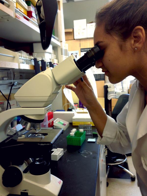
Each bench was covered in scientific tools including pipets ranging from different sizes, flasks and beakers filled with chemicals, slides stained with different specimens, elaborate microscopes, and measuring equipment. The room itself contained other smaller rooms in which the more expensive equipment was located, including compound light microscopes, the scanning probe microscope and the electron microscope.
I was assigned two mentors, Crista Grant and Ryan Spurrier — both research fellows working towards their goals of becoming surgeons. Throughout my first week, they gave me a rundown of what their lab does in relation to the small intestine and Short Bowel Syndrome.
After numerous explanations and clarifying of scientific jargon, I finally had a basic understanding of the illness and the research conducted at the lab. I learned that Short Bowel Syndrome is caused by a shortage in the small intestine, which prevents its victims from properly absorbing nutrients. Crista and Grant told me about various somber cases of children with this illness and the trouble they and their families encountered. These children spend each day trapped within the white hospital room walls, suffering from various health implications including fatigue, vomiting and weight loss. This illness has taken away the lives of thousands of children and troubled their families who not only watch their kids in agony but also are burdened with costly medical bills.
Dr. Grikscheit’s lab takes a novel approach to treating this illness by using stem cells to produce tissue-engineered small intestine. “The ultimate goal of this lab,” said Grant, “is to save the lives of babies, who would otherwise die because they had their intestine cut out by surgery.”
I came into this lab thinking it was all about the science and facts, but hearing these stories added a sentimentality to the work and made me passionate about taking part, although my role was minor, just a small part of a larger and greater picture.
The first machine I learned to use was the microtome, which is used to section blocks of tissue fixed in formalin (wax). This immense machine used a sharp blade, which I managed to cut my finger on, to cut thin pieces of tissue, which would later be observed. Although it serves a basic function, this machine is actually a lot more difficult to use than it seems. Using it required a precise method and certain delicacy, which I did not have.
Adding to my frustration, the thin pieces of tissue kept ripping from the force of my hand, making them useless. In the beginning, I would discard about 70% of my block. After finally cutting the tissue, I would put the pieces in a dish of warm water to eliminate creases in the wax. I then encountered the problem of transferring these sections from the water dish to a slide.
It took me a few weeks to finally master the art of sectioning blocks and developing my own technique. My initial feeling of frustration did not last very long, as I grew to love this process because it required a certain concentration and art, which I found it relaxing.
The next skill I learned in the lab was the art of staining. There are two types of staining: H&E and immunoflourescent staining. The former is the standard stain in histology, which stains for general parts of cells such as the nucleus and the latter stains for specific proteins. In both stains, I had to place slides in a succession of dishes, each with different colored dyes. The difference between each process is the amount of time the slides are left inside the dish and which specific dies they were placed in.
The process itself was not difficult; however, it required me to be extremely meticulous because I had to time how long the slides remained inside the dyes. This was definitely a challenge for me because I do not pay attention to small details, and often times I would leave the slides in for too long in a certain dye and have to start the whole process over again.
After I stained the slides, I would accompany Crista and Ryan in observing them under the microscope and deciphering what the slides portrayed and proved. We would then capture images of the slides, which would later be used in scientific reports.
I was even fortunate enough to participate in Ryan’s experiment. My role involved comparing how closely the tissue-engineered intestine resembled a native intestine by staining for the different specialized stem cells in both small intestine, capturing images through the microscope, and documenting what corresponded. The results of the experiment were remarkable, as the two were extremely similar. According to Ryan, “We proved what the whole goal of this lab was.”
I definitely made my fair share of mistakes in the lab and had to discard various materials or redo several processes. I often grew nervous because the work I was producing was not just for practice. It was actually going to be used by Crista and Ryan in their experiments.
I was definitely feeling the pressure and would sometimes feel discouraged; however, I was fortunate to have very understanding mentors who did not mind repeating themselves various times and helping me master the basic skills used in the lab through demonstrations and thorough explanations. What made my experience so special was that the members of the lab themselves were close knit and made me feel like part of the family. It was easy for me to become comfortable around them and form close relationships.
My experience at the lab opened my eyes up to the vitality of progressing science. According to Crista, “Science at the bench requires a lot of techniques to learn about such as PCR. In the beginning it’s really hard. There’s surgical techniques to learn, and of course the challenge of proving your hypothesis, which is that the intestinal stem cell can drive regeneration of a new small intestine. We have to figure out and design an experiment to prove that. The ultimate goal of this lab is to save the lives of babies, who would otherwise die because they had their intestine cut out by surgery.”
During my second time in the conference room, those same doctors who were fiercely debating on the science of the cells of one patient, were now discussing another patient’s death and her family’s difficulty in coping. Their words which once sounded so robotic and isolating were now understandable and relatable to. To many on the outside, science and lab work appear to be based on the cold hard facts, and although that is true, they also have a very human appeal and evoke pathos. During my experience I came to understand how this dichotomy harmoniously exists in a lab.


